Loculated Pleural Effusion Cxr / Loculated Pleural Effusion Radiology Case Radiopaedia Org - Pleural effusions are a common medical problem with more than 50 recognised causes including disease local to the pleura or underlying lung, systemic conditions, organ dysfunction and drugs.1.
Loculated Pleural Effusion Cxr / Loculated Pleural Effusion Radiology Case Radiopaedia Org - Pleural effusions are a common medical problem with more than 50 recognised causes including disease local to the pleura or underlying lung, systemic conditions, organ dysfunction and drugs.1.. Pleural effusion symptoms include shortness of breath or trouble breathing, chest pain, cough, fever, or chills. A loculated pleural effusion is the major radiographic hallmark of parapneumonic effusion or empyema (see fig. Most commonly caused by a viral infection. What does pleural effusion mean? Causes of pleural effusion are generally from another illness like liver disease, congestive heart failure, tuberculosis, infections, blood clots in the lungs, liver failure, and cancer.
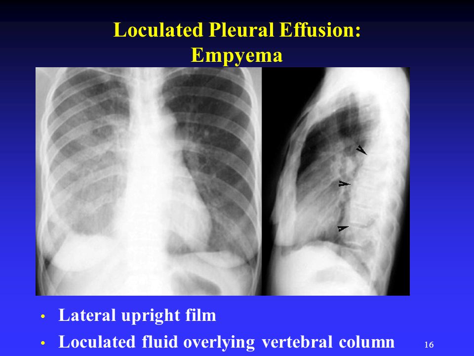
Pleural fluid ldh > two thirds of upper limit for serum ldh. What does pleural effusion mean? Pleural fluid/serum protein ratio >0.5. Pleural fluid/serum ldh ratio >0.6. Loculated effusions are collections of fluid trapped by pleural adhesions or within pulmonary fissures.
Pleural Disease Ppt Video Online Download from slideplayer.com This is typical of a pseudotumor due to a loculated pleural effusion distending the transverse fissure. Pleural fluid/serum ldh ratio >0.6. What does pleural effusion mean? Not respond to chest tube and antibiotics. Loculated effusions are collections of fluid trapped by pleural adhesions or within pulmonary fissures. Other causes are complicated parapneumonic effusion. Approximately 1 million people develop this abnormality each year in the united states. Pf ada levels, nodular lung lesions, and loculated pleural effusion may help differentiate tpe from ppe in patients with pf showing.
Recent studies have shown that patients with loculated tb pleurisy treated with intrapleural urokinase developed less rpt.
The pleura are thin membranes that line the lungs and the inside of the chest cavity and act to lubricate and facilitate breathing. Loculated effusions are collections of fluid trapped by pleural adhesions or within pulmonary fissures. Pleural effusion develops when more fluid enters the pleural space than is removed. Pleural effusions are diagnosed in about 1.5 million individuals in the united states annually. Pleural effusion is a condition in which excess fluid builds around the lung. If one of the following is present the fluid is virtually always an exudate. Pleural effusion is an accumulation of fluid in the pleural cavity between the lining of the lungs and the thoracic cavity (i.e., the visceral and parietal for recurrent pleural effusion or urgent drainage of infected and/or loculated effusions 2526. Approximately 1 million people develop this abnormality each year in the united states. Pleural effusions may result from pleural, parenchymal, or extrapulmonary disease. If none is present the fluid is virtually always a transudate. Always do pleural biopsy if you suspect tb.disorder in the workup of a pleural effusion after performing thoracentesis always order. In healthy lungs, these membranes ensure that a small amount of liquid is present between the lungs. Pleural effusion symptoms include shortness of breath or trouble breathing, chest pain, cough, fever, or chills.
If none is present the fluid is virtually always a transudate. Approximately 1 million people develop this abnormality each year in the united states. Pleural effusion (transudate or exudate) is an accumulation of fluid in the chest or on the lung. Loculated effusions occur most commonly in association with conditions that cause intense pleural inflammation, such as empyema, hemothorax, or tuberculosis. Commonly from congestive heart failure or malignancy.
Pdf Intrapleural Streptokinase For Tuberculosis Loculated Pleural Effusion Semantic Scholar from d3i71xaburhd42.cloudfront.net This is typical of a pseudotumor due to a loculated pleural effusion distending the transverse fissure. There is a large left pleural effusion obscuring the lower half of the left hemi thorax. Most commonly caused by a viral infection. When you have a pleural effusion, fluid builds up in the space between the layers of your pleura. Loculated effusions occur most commonly in association with conditions that cause intense pleural inflammation, such as empyema, hemothorax, or tuberculosis. What does pleural effusion mean? Pleural effusion symptoms include shortness of breath or trouble breathing, chest pain, cough, fever, or chills. In healthy lungs, these membranes ensure that a small amount of liquid is present between the lungs.
Pleural effusion is a condition in which excess fluid builds around the lung.
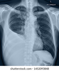
There is a large left pleural effusion obscuring the lower half of the left hemi thorax. Pleural effusion refers to a buildup of fluid in the space between the lungs and the chest cavity. Treatment depends on the cause. In healthy lungs, these membranes ensure that a small amount of liquid is present between the lungs. Always do pleural biopsy if you suspect tb.disorder in the workup of a pleural effusion after performing thoracentesis always order. Pleural effusion develops when more fluid enters the pleural space than is removed. The cardiac silhouette is also obscured. Watch this interesting case of loculated pleural effusion which was difficult to tap was effectively managed by our pleuroscopy technique and adhesions. The cxr shows classic evidence of congestive heart failure with cardiomegaly, upper lobe venous diversion, and bilateral pleural effusions. Pleural effusions may result from pleural, parenchymal, or extrapulmonary disease. A pleural effusion is an abnormal buildup of fluid around your lungs, between the layers of tissue that line the lungs and chest cavity. Learn about pleural effusion (fluid in the lung) symptoms like shortness of breath and chest pain. Pleural fluid/serum ldh ratio >0.6.
A pleural effusion is accumulation of excessive fluid in the pleural space, the potential space that surrounds each lung. Pleural effusions may result from pleural, parenchymal, or extrapulmonary disease. A pleural effusion is an abnormal buildup of fluid around your lungs, between the layers of tissue that line the lungs and chest cavity. More than one half of these massive pleural effusions are caused by malignancy; Loculated effusions occur most commonly in association with conditions that cause intense pleural inflammation, such as empyema, hemothorax, or tuberculosis.
Loculated Pleural Effusion Images Stock Photos Vectors Shutterstock from image.shutterstock.com How is pleural effusion detected. Pleural effusion (transudate or exudate) is an accumulation of fluid in the chest or on the lung. A pleural effusion is accumulation of excessive fluid in the pleural space, the potential space that surrounds each lung. The cardiac silhouette is also obscured. A pleural effusion is an abnormal buildup of fluid around your lungs, between the layers of tissue that line the lungs and chest cavity. Send aspirated fluid for cytology. Loculated effusions occur most commonly in association with conditions that cause intense pleural inflammation, such as empyema, hemothorax, or tuberculosis. Meaning of pleural effusion medical term.
Commonly from congestive heart failure or malignancy.
Pleural fluid/serum ldh ratio >0.6. Loculated pleural effusion on cxr. Pleural fluid/serum protein ratio >0.5. Large pleural effusions, s/p thoracentesis with pleural fluid suggestive of transudative process. Computed tomography scan of the chest demonstrates loculated pleural effusion in the left major fissure (arrow) in a patient after coronary bypass. Pleural fluid ldh > two thirds of upper limit for serum ldh. Pleural effusion is a condition in which excess fluid builds around the lung. When you have a pleural effusion, fluid builds up in the space between the layers of your pleura. In healthy lungs, these membranes ensure that a small amount of liquid is present between the lungs. Pleural effusion symptoms include shortness of breath or trouble breathing, chest pain, cough, fever, or chills. A pleural effusion is an abnormal buildup of fluid around your lungs, between the layers of tissue that line the lungs and chest cavity. The pleura is a thin membrane that lines the surface of your lungs and the inside of your chest wall. Always do pleural biopsy if you suspect tb.disorder in the workup of a pleural effusion after performing thoracentesis always order.
The pleura are thin membranes that line the lungs and the inside of the chest cavity and act to lubricate and facilitate breathing loculated pleural effusion. Loculated pleural effusion on cxr.


0 Komentar